
Novel 4D MRI Method for In Utero Imaging
University of Pittsburgh and Cedars-Sinai Medical Center scientists have developed a novel magnetic resonance imaging (MRI) method to study and simultaneously assess fetal and placental development, morphology, and functions. This approach uses a combination of conventional MRI (cMRI) and functorial MRI (fMRI) captured at high frame rates (>60 frames per second (fps)) together with novel specialist “image-reconstruction” software to visualize both anatomy and function of a fetus (or multiple fetuses) in utero using a single scan. This novel 4D MRI method can provide previously unobtainable levels of detail of cardiac and brain imaging and could revolutionize the field of fetal medicine.
Description
In utero fetal imaging is vital to assist the diagnosis and treatment planning for cardiac or brain anomalies discovered during pregnancy. Congenital heart defects (CHD) affect nearly 1% of live births, making CHD the most common birth defect. While surgery has improved outcomes in these children, comorbidities are common including neurocognitive impairment. The underlying cause of these comorbidities could be intrauterine hypoxia, placental structure, vascular abnormalities, or a combination of these. Current research techniques have failed to definitively reveal the relationship between fetal CHD and neurocognitive impairment. This novel MRI method would allow for in-depth studies of animal models of CHD and other developmental defects in utero leading to new advances in fetal medicine including treatment plans ultimately improving outcomes in patients with some congenital defects.Applications
• Fetal medicine research• Congenital defect screen in pregnancy
Advantages
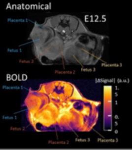
MRI, both conventional and functional, is used for some fetal imaging. MRI fails to fully visualize many of the dynamic functions of the placenta and fetus due to issues including fetal and maternal motions and insufficient frame rates.This novel technique overcomes the current shortcomings of MRI by combining cMRI with the high frame rate of fMRI. Full 3D isotropic morphological imaging for multiple fetuses and placentas simultaneously, with imaging of tissue functions including cardiac and hemodynamic functions, is obtained from a single scan. Using a frame rate over 60 fps reduces image artifacts from motion, improving visualization quality and accuracy. Images are reconstructed using custom software and training data to decode spatially encoded data. This 4D MRI method can be used on commercially available MRI scanners with only minor software alterations required. With development, this technique could be used in gestational screening.
Invention Readiness
In utero 4D MRI was performed in a pregnant mouse on embryonic day E16.5. Isotropic 3D data of eight embryos were obtained, including detailed images of brain structures.IP Status
https://patents.google.com/patent/US11828825B2Related Publication(s)
Reynolds, W. T., Votava-Smith, J. K., Gabriel, G., Lee, V. K., Rajagopalan, V., Wu, Y., Liu, X., Yagi, H., Slabicki, R., Gibbs, B., Tran, N. N., Weisert, M., Cabral, L., Subramanian, S., Wallace, J., Del Castillo, S., Baust, T., Weinberg, J. G., Lorenzi Quigley, L., Gaesser, J., … Lo, C. W. (2024). Validation of a Paralimbic-Related Subcortical Brain Dysmaturation MRI Score in Infants with Congenital Heart Disease. Journal of clinical medicine, 13(19), 5772. https://doi.org/10.3390/jcm13195772